TABLE OF CONTENTS
Anatomical Classification of Placenta
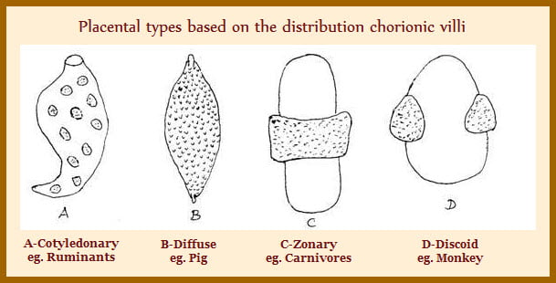
Anatomical classification of placenta is based on chorionic villi distribution in the placenta. It may be diffuse, cotyledonary, zonary, or discoidal placenta.
Placenta is divided in to 4 general types based on their shape as:
- Diffuse
- Cotyledonary
- Zonary
- Discoidal
1. Diffuse
The diffuse placenta of the sow consists of numerous chorionic villi on surface of chorion. They penetrate into endometrium forming fetomaternal interface.
The diffuse placenta of the mare has many micro-cotyledons on the surface of the chorion, which are responsible for fetal-maternal exchange.
The mare placenta is characterised by having numerous specialized microzones of chorionic villi known as microcotyledons. These are the discrete regions at fetal maternal interface. Mare placenta also contains transitory structures known as endometrial cups. It is of both endometrial and trophoblastic origin.
There were 5-10 endometrial cups distributed over the surface of the placenta which secrete equine chorionic gonadotropin (eCG). And develop between days 35-60 of pregnancy. Following day 6o the endometrial cups are sloughed into uterine lumen and no longer functional.
2. Cotyledonary
Ruminants have cotyledonary placenta. A cotyledon is defined as placental unit of trophoblastic origin consisting of abundant blood vessels and connective tissue. In sheep there are between 90-100 cotyledons distributed across the surface of the chorion and in cattle around 70-120 cotyledons have been observed.
The placentome (point of interface) In the cotyledonary placenta consist of fetal cotyledon contributed by chorion and a maternal cotyledon originating from the caruncular regions of the uterus. At about day 16 in sheep and day 25 in cow the chorion initiates attachment to the caruncles of the uterus.
Prior to this time the placenta is essentially diffuse. During the formation of placentome, chorionic villi protrude into crypts in the caruncular tissue. Attachment is well established by day 30 in ewes and day 40 in cows.
Cotyledonary placentas are char by button like structures on the surface of the chorion. They are referred as fetal cotyledons. They join with maternal caruncle and form a placentome.
A convex cotyledon becomes covered with chorion. Many finger like villi originating from the chorionic tissue protrude toward the lumen of the uterus. In the concave cotyledon, the chorionic tissues push inward, forming a concave interface between the chorion and maternal caruncle.
3. Zonary
The zonary placenta includes a prominent region of exchange that forms a broad zone around the chorion near the middle of the conceptus.
A second region consists of highly pigmented ring at either end of the central zone. The pigmented zone consists of small hematomas.
The pigmented zone is also referred as paraplacenta and is thought to be important in iron transport from the dam to fetus. The function of this zone is not well understood.
A third region is the transparent zone on the distal ends of the chorion that has poor vascularity. This zone may be involved in absorption of materials directly from the uterine lumen.
4. Discoidal
The discoid placenta is found in rodents and primates. It is characterised by having one or two distinct adjacent discs.
These discs contain chorionic villi that interface with endometrium and provide the region for nutrient and metabolic waste exchange.

