TABLE OF CONTENTS
Affections of Kidney and urinary system
Affections of Kidney and urinary system are Ectopic ureter, Uroabdomen or Uroperitoneum, Urolithiasis, Vesical Calculi, Patent Urachus, Urethral Calculi and Stenosis, Urethral prolapse etc.
Surgical affections of Kidney, ureter, urinary bladder and urethra in animals are-
Surgical procedures of Kidney, ureter, urinary bladder and urethra in animals are-
Ectopic ureter
Ectopic ureter is a congenital anomaly where one or both ureters empty outside the urinary bladder.
Symptoms
- Urinary incontinence
- Pyelonephritis and cystitis
- Hydroureter
- Suspect Ectopia of ureter in any young animal that has a history of incontinence since birth
- Suspect this in older animals with lifelong urinary incontinence
- More commonly diagnosed in female dogs than in males
Diagnosis
- Contrast Radiography
- Ultrasonography
Treatment
Medical management aimed at treating the urethral sphincter incompetence. Drugs used are-
- Phenylpropanolamine
- Ephedrine
- Imipramine
- Diethylstilbestrol
Surgical techniques
Ectopic ureter includes the following surgical techniques-
- Neoureterostomy
- Ureteroneocystostomy
Neoureterostomy
Neoureterostomy is performed for intramural ectopic ureters where the ureter course submucosally in the bladder and opens into the urethra or vagina. After performing a cystotomy, an incision is made into the ureter to make a stoma into the bladder. This is followed by ligation of the ureter coursing distally to the stoma.
Ureteroneocystostomy
Ureteroneocystostomy is performed for extraluminal ectopic ureters where the ureter completely bypasses the bladder. The ureter is first ligated and transected. An new entry for the ureter is then created in the bladder and the transected end of the ureter is then sutured to the bladder mucosa through the newly created bladder incision.
Uroabdomen or Uroperitoneum
Uroabdomen or Uroperitoneum is a condition where there is accumulation of urine in the peritoneal cavity due to leakage of urine from the kidney, ureter, bladder or proximal urethra.
Rupture of the bladder is the most common cause. Also due to blunt or penetrating trauma (examples are Automobile accidents, Penetrating injury from the fracture fragments of pelvic bone).
Remember that if the rupture of the bladder is small or on its dorsum, leakage will happen only when the bladder is fully distended. In such cases, the dog may void urine normally.
Diagnosis
- Suspicion from history of trauma
- Peritoneocentesis reveal smell of urine in the fluid and the creatinine level will be greater than that of serum
- From physical examination findings
- Confirmatory diagnosis are Plain or Contrast radiography and Ultrasonography
Medical management
If the animal is hyperkalemic or uremic, medical treatment should be initiated. Peritoneal dialysis is preferred in such situations.
Surgical treatment
- Repair trauma of the urethra by anastamosis
- If the urethra is not completely transected, it can be allowed to heal over a urinary catheter
- Rupture of the ureter can be corrected either by anastamosis or by reimplantation into the bladder
- Rupture of the bladder (mostly at the apex) can be corrected by apposition of the edges.
Urolithiasis
Urolithiasis is the Renal, Ureteral, Cystic or Urethral calculi.
Urolithiasis means a condition of having urinary calculi or uroliths. Can be any of the following-
- Nephrolith– calculi in the kidney
- Ureterolith– calculi in the ureter
- Cystolith– calculi in the urinary bladder
- Urethrolith– calculi in the urethra
Symptoms
- Some breeds have higher incidence due to metabolic abnormalities (eg. Dalmatians)
- Signs depends on whether the stone has caused obstruction or associated infection is present
- Renal calculi cause hematuria, flank pain or renomegaly
- Polyuria and polydipsia if the animal has pyelonephritis
Diagnosis
- From symptoms
- Radiography
- Ultrasonography
Treatment
- Treat the underlying infection
- If the calculi is associated with obstruction, surgical removal is essential Surgical removal include the following
- Nephrotomy for removal of renal calculi
- Cystotomy for cystoliths
- Urethrotomy for calculi in the urethra
Vesical Calculi
Vesical calculi are stones or calcified materials that are present in the urinary bladder. Symptoms related to stones in the urinary bladder are not always shown.
Chronic cases will exhibit hamaturia and dysuria. Sometime complete obstruction of the urethra with stones in male dogs will cause bladder distension.
In female there may not be bladder distension, but soiling of the perineal region with urine and urine smell will be there., palpation reveals crepitation of the calculi in the bladder.
Diagnosis
Diagnosis is based on symptoms, and palpation of the posterior abdomen will reveal stone like bladder.
Radiography
Plain radiography will reveal distended bladder and vesical claculi if radiopaque will reveal stones. If they are radiolucent, pneumocystography will confirm the diagnosis.
Treatment
Cystotomy is the only surgical treatment in case of Vesical Calculi.
Surgical procedure
- Place the animal in dorsal recumbency
- Prepare ventral abdominal region and vulvar area in female for aseptic surgery
- Incise skin and subcutis on the ventral midline
- In male incise skin and subcutis parallel and adjacent to prepuce
- Identify and ligate preputial branches of caudal superficial epigastric artery in the subcutis
- Incise linea alba from umbilicus to pubis and para preputial approach in male dogs
- Identify bladder and isolate it by moistened laparotomy sponges.
- Place stay sutures on the bladder apex to facilitate manipulation
- Make incision on the dorsal aspect of bladder away from ureters , urethra and between major blood vessels
- Remove urine by aspiration or intraoperative cystocentesis before cystotomy.
- remove the cystoliths
- Flush the bladder with normal saline to remove small calculi
- Examine bladder and mucosa for defects
- Pass a catheter down the urethra to check for patency
- Close the urinary bladder (Cystorraphy)in a single layer using continuous suture pattern
- If two layer closure, suture the seromuscular layer by two continuous inverting suture lines (cushings suture followed by lembert)
Patent Urachus
Patent urachus refers to umbilical disorders that result from the failure of involution of normal embryologic tissues that connect the developing bladder to the umbilical cord.
Patent urachus is a common condition in calves which is frequently associated with omphalitis. A membranous urethral diaphragm prevented closure of the urachus in a female calf. The patent urachus was complicated by an ascending infection of the intra-abdominal umbilical remnants.
Urethral prolapse
Urethral prolapse is the prolapse of the mucosal lining of the distal portion of the urethra through the external urethral orifice, common in english bull dogs.
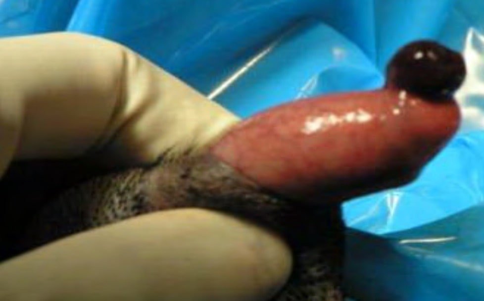
- Reddened protrusion at the tip of the penis
- Sometimes it may be seen intermittently when the dog is sexually excited o Some dogs lick and traumatize the prolapse, and it may bleed
Symptoms
- persistent licking of the penile area
- red to purple pea sized lesion
- hematuria independent of micturition
Management
- May resolve spontaneously if the size is small
- Manual reduction under general anaesthesia using a urinary catheter, purse string suture is applied and later removed after 4 days
- Resection of the prolapsed mass is carried out in extensively damaged cases
Treatment
- If the prolapse is reducible, reduction followed by retention with sutures from the urethral lumen to the penile surface can be done
- Surgical resection of the prolapse is the choice when the prolapse cannot be reduced
Cystotomy
Cystotomy is a surgical incision into the urinary bladder.
Indications
- Cystic calculi
- Neoplasia of urinary bladder
- Correction of ectopic ureters
- Urinary tract infection which is resistant to treatment
Symptoms
- Typical symptoms include straining to urinate (stranguria)
- Blood in the urine (hematuria)
- Urinating small amounts frequently (pollakiuria)
- Excess urination (polyuria)
- Pain in the rear quarters
- Reluctance to jump or play, or even lethargy
Diagnosis
- A urinalysis is helpful in making a diagnosis. The pH of the urine, mineral content, and the presence of bacteria or crystals all provide valuable information
- Radiography-radiopaque calculi can be detected
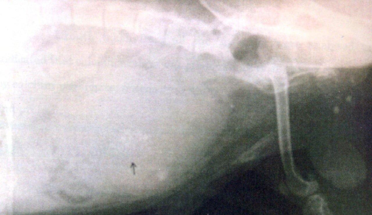
Ultrasonography is a good method to diagnose stones in the urinary bladder, particularly for radio lucent calculi and anatomical defects of the abdominal wall.
Pre operative care
Correct the fluid and electrolyte imbalances because of chances of hyperkalemia associated with urinary obstruction.
Surgical procedure
- Place the animal in dorsal recumbency
- Prepare ventral abdominal region and vulvar area in female for aseptic surgery
- Incise skin and subcutis on the ventral midline
- In male incise skin and subcutis parallel and adjacent to prepuce
- Identify and ligate preputial branches of caudal superficial epigastric artery in the subcutis
- Incise linea alba from umbilicus to pubis and para preputial approach in male dogs
- Identify bladder and isolate it by moistened laparotomy sponges
- Place stay sutures on the bladder apex to facilitate manipulation
- Make incision on the dorsal or ventral aspect of bladder away from ureters, urethra and between major blood vessels and remove the calculus with a forceps
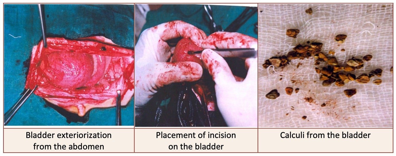
- Remove urine by suction or intraoperative cystocentesis before cystotomy
- Examine bladder and mucosa for defects
- Pass a catheter down the urethra to check for patency
- Close the UB in a single layer using continuous suture pattern
- If two layer closure, suture the seromuscular layer by two continuous inverting suture lines (cushings followed by lembert)
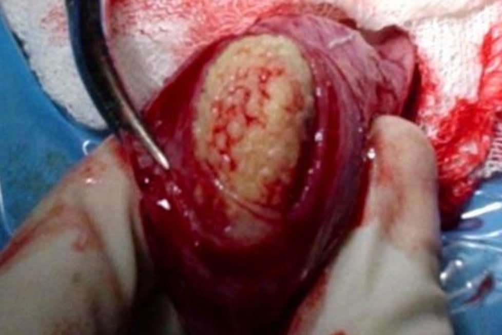
Post operative care
- Antibiotic and NSAIDS
- Observe the site twice daily for redness, swelling or discharge from the site and cleaning of the surgical site
- Suture removal after 10-12 days
- Special diet recommendations based on the type of calculi
- If struvite-decreased protein, give acidifiers such as ascorbic acid and dl-methionine
- If calcium oxalate, decrease protein, calcium, sodium (spinach, milk products, table salt)
- If urate, increase the water consumption, feed a diet low in purines, increase in pH 7.0-7.5 (Potassium citrate), adding allopurinol to prevent conversion of purine to uric acid
Cystorrhaphy
Suturing of a wound due to injury or rupture of urinary bladder. Suture seromucosal layer with two continuous inverting suture lines (Cushings followed by Lembert)
If bladder wall is thickened, suture bladder using a continuous suture pattern using absorbable suture material
If the dog has severe bleeding tendencies, suture mucosa as a separate layer with a simple continuous suture pattern
Urethrotomy
Urethrotomy is a surgical procedure used to remove urethral calculi most frequently in male and occasionally in female when hydro-propulsion fails to flush the calculi into the bladder.
A urethrotomy is a surgical operation to treat a narrowing of your urethra.
Indication
- Urethral calculi
- Prostate diseases
- Urethral obstruction
- Occasionally, biopsy of obstructive lesions (i.e. strictures, scar tissue and neoplasms)
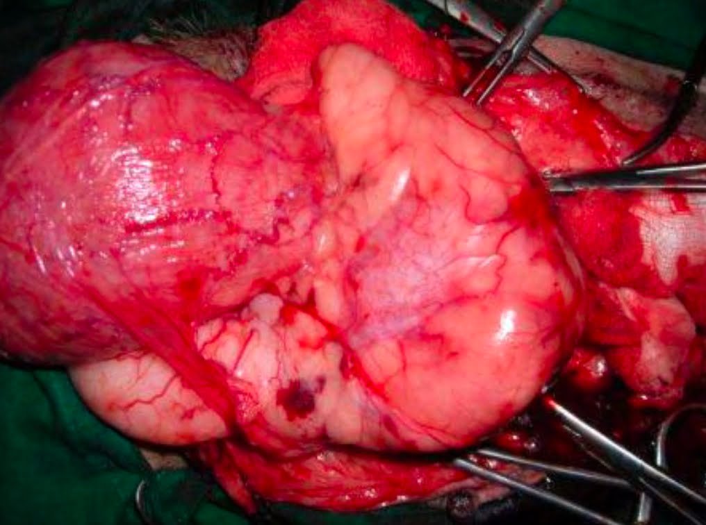
Incision site
- Bull– 3 sites-
- Post scrotal site: For removal of obstruction at the sigmoid flexure, about 3 inches behind the scrotum along the median line.
- Ischial site: For obstruction close to the ischial arch. Two inches below the ischial arch downwards along midline.
- Ventral approach: Between scrotum and preputial orifice 5-6 inch incision over the midline centering the lodged calculi.
- Horse– The median line of perineal region, at or below the level of ischial arch
- Dog– At the seat of obstruction (the commonest site of obstruction is behind the os-penis and occasionally calculi lodged at the ischial arch)
- Sheep & Goat– The urethral calculi in rams and wethers are mostly lodged in the urethral process or in the sigmoid flexure
Operation technique in Bulls
Post scrotal method
- A midline incision of 3 inches long is made about 3 inches behind the scrotum.
- The areolar tissue is dissected to reveal retractor penis muscles on either side.
- These are separated and held retracted to expose the body of the penis.
- Palpate the urethra on the ventral aspect and incise it longitudinally along the exact midline.
- The blockage is relieved and the patency of the canal is established by a gum elastic catheter or a pliable metal probe.
- A thin elastic tube may be used as a catheter and left in situ for one or two days.
- The wound is left open to heal by second intension which ensures if the normal passage is clear.
Ischial urethrotomy
- A skin incision 2 inches long is made along the midline starting from about 2 inches below the ischial arch downwards. This exposes the two retractor penis muscles.
- The incision can be started in level with the ischial arch but will cause unnecessary bleeding due to the cutting of the ischeo-cavernous muscle.
Operation technique in Dogs
Pre-scrotal urethrotomy
- With the dog in dorsal recumbency, place a sterile catheter into the penile urethra to the scrotum or to the obstruction.
- Make a ventral midline incision through the skin and subcutaneous tissue between the caudal aspect of the os penis and scrotum.
- Identify, mobilize and retract the retractor penis muscle laterally to expose the urethra.
- Using a scalpel blade, make an incision into the urethral lumen over the catheter.
- Use iris scissors to extend the incision, if necessary.
- Remove calculi with forceps and gently flush the urethra with warm saline.
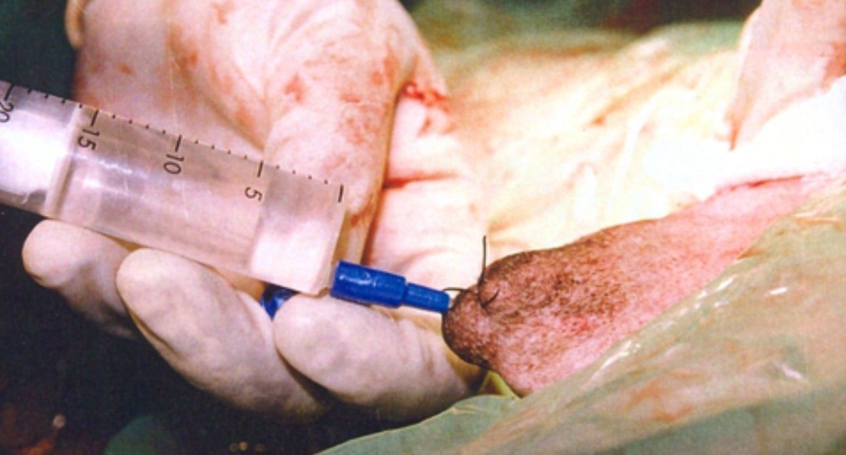
Leave the incision to heal by secondary intention or close the urethra with simple interrupted absorbable sutures (4-0 or 5-0)
Place the first layer in the urethral mucosa and corpus spongiosum then appose subcutaneous tissue and skin with simple interrupted sutures or a continuous subcuticular suture pattern
Perineal urethrotomy
- Place a purse string suture in the anus.
- Place a sterile catheter into the urethra to the level of the bladder or the site of the obstruction
- With the dog in sternal recumbency and rear limbs hanging over the edge of the table, make a midline incision over the urethra, midway between the scrotum and anus
- Identify the retractor penis muscle, elevate it and retract it
- Separate the paired bulbospongiosum muscles at their raphe to expose the corpus spongiosum then incise the corpus spongiosum to enter the urethral lumen
- Close the incision as just described for prescrotal urethrotomy
Operation technique in stallion
- A median cutaneous incision is made in the perineal region at the ischial arch, 3 to 4 inches long
- Go between the retractor penis muscles and cut through the accelerator urinae muscle, corpus spongiosum and the urethral wall.
- Confine to the exact median line to avoid branches of the internal pudic artery
- The wound may be left open or alternatively the urethra may be sutured to correspond to the skin edges to keep the opening patent
Operation technique in Sheep and Goats
- The animal is restrained; the penis is pulled out gently from the prepuce and digital pressure applied on the urethral process to remove the calculus.
- In wethers, alternatively, urethral process can be nicked off with a scissors and minor bleeding checked with digital pressure.
- The technique of post scrotal urethrotomy to remove a calculus from the sigmoid flexure is same as described in bovines.
Post Operative care
- The animal should receive adequate fluid therapy immediately after surgery and for a few days afterwards to correct hypovolaemia.
- Analgesics should be administered fore 3 to 4 days and broad spectrum antibiotics are given for 5 to 7 days to check secondary infection.
- Routine wound dressing should be done daily.
- The indwelling catheter is left in situ for 3 to 4 weeks.
- Corticosteroids are often used for 2 to 3 days.
- The animal should be carefully watched for any complications, particularly for subcutaneous infiltration of urine due to its seepage from the urethral wound.
- Administer orally Cystone ® tablets which are thought to act as urinary antiseptic and also to avoid recurrence of calculus formation.
Complications of urethrotomy
- Blockade of the urethral catheter occurs mostly due to blood clot and casts or renal or cystic cells, kinking of the catheter and apposition of the proximal rim of the catheter against the urethral wall
- Urethral wound dehiscence may occur due to infection or seepage of urine.
- Urethral stricture or urethral stenosis
- Primary atony of the bladder occurs rarely in cattle and buffaloes. It may develop secondary to obstructive urolithiasis due to over distension or as a complication of bladder surgery
- Peritonitis is a rare post-operative complications